Do Mini Strokes Show Up on CT Scans? Understanding the Imaging of Transient Ischemic Attacks
When a person experiences a sudden, brief neurological deficit, doctors often suspect a transient ischemic attack (TIA), commonly called a “mini‑stroke.” One of the first questions patients ask is whether a CT scan can detect the event. The short answer is that most TIAs do not produce visible changes on a standard non‑contrast CT scan, but imaging still plays a crucial role in ruling out other causes and guiding treatment.
What Is a Mini Stroke?
A mini stroke, or TIA, occurs when blood flow to a part of the brain is temporarily reduced or blocked, usually lasting less than 24 hours. Unlike a full‑blown ischemic stroke, the symptoms—such as weakness, speech difficulty, or vision loss—typically resolve completely within minutes to hours. Because the brain tissue often recovers without permanent injury, the term “mini stroke” can be misleading; the underlying vascular problem may still be serious.
Why CT Scans Are Frequently Ordered
Emergency departments rely on computed tomography (CT) for several reasons:
- Speed: A head CT can be completed in under five minutes, providing rapid information when time is critical.
- Exclusion of hemorrhage: CT is highly sensitive to acute bleeding, which must be ruled out before administering antiplatelet or anticoagulant therapy.
- Availability: Most hospitals have CT scanners on standby 24/7, whereas MRI may have limited access.
These advantages make CT the first‑line imaging test for anyone presenting with stroke‑like symptoms, even when a TIA is suspected.
What a Standard CT Scan Typically Shows
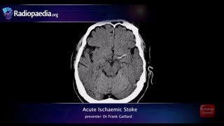
In the majority of TIAs, a non‑contrast CT scan appears normal. The brief interruption of blood flow usually does not cause enough tissue damage to create the radiodense changes that CT detects. As Dr. Erik Pedersen explains in his introductory lectures on neuroimaging, “CT is excellent for spotting blood, but subtle ischemic changes often require more sensitive modalities.”
However, there are exceptions:
- Large‑vessel TIAs: When a major artery such as the middle cerebral artery is briefly occluded, a small area of early ischemia may be visible as a faint hypodensity.
- Posterior circulation events: The brainstem and cerebellum are harder to assess on CT, and sometimes a subtle loss of gray‑white differentiation can be seen.
- Concurrent pathology: A CT may reveal a tumor, aneurysm, or subdural hematoma that mimics TIA symptoms.
When MRI Becomes the Preferred Tool
Magnetic resonance imaging (MRI), especially diffusion‑weighted imaging (DWI), can detect ischemic changes within minutes of onset. Studies referenced in the AJR article by Andrew Rosenkrantz, MD demonstrate that DWI MRI identifies lesions in up to 70 % of patients with clinically diagnosed TIAs, compared with less than 10 % on CT.
Linda Ciampa, a neurologist who frequently discusses TIAs, emphasizes that “MRI is the gold standard for confirming a mini stroke, but accessibility and cost often dictate initial CT use.” When a CT scan is normal but suspicion remains high, clinicians usually order an MRI to look for the tiny infarcts that CT missed.
How Imaging Influences Management
Even a normal CT scan provides valuable information:
- Rule out hemorrhage: Allows safe initiation of antiplatelet therapy.
- Identify alternative diagnoses: Detects masses, infections, or structural abnormalities that require different treatment.
- Guide further testing: A normal CT may prompt carotid ultrasound, cardiac monitoring, or MRI to uncover the source of the TIA.
Guidelines from the American Heart Association recommend that patients with a suspected TIA receive both a CT (or MRI) and vascular imaging (CTA, MRA, or carotid duplex) within 24 hours to assess stroke risk and determine the need for interventions such as carotid endarterectomy.
Practical Takeaways for Patients
If you experience sudden neurological symptoms that resolve quickly, seek emergency care immediately. Expect the following pathway:
- CT scan on arrival: Primarily to exclude bleeding.
- Neurological exam: Determines the likelihood of a TIA.
- Additional imaging (MRI, CTA): Ordered if the CT is normal but clinical suspicion persists.
- Risk‑reduction plan: May include antiplatelet medication, blood pressure control, cholesterol management, and lifestyle changes.
Remember that a normal CT does not guarantee that a mini stroke didn’t occur; it simply means no acute bleed or large‑scale damage was seen. Follow‑up with your physician is essential to address underlying vascular risk factors.
Where to Find More Reliable Information
For a deeper dive into stroke and TIA education, visit reputable resources such as Khan Academy’s health and medicine section. Their videos break down complex topics into easy‑to‑understand lessons, covering everything from blood flow dynamics to preventive strategies.
Conclusion
In summary, mini strokes (TIAs) rarely show up on standard CT scans, but the imaging is still a critical first step in the emergency evaluation. A normal CT helps rule out hemorrhage and other serious conditions, while MRI and vascular studies provide the sensitivity needed to confirm an ischemic event and guide long‑term prevention. Prompt medical attention, appropriate imaging, and diligent follow‑up remain the best defenses against future strokes.